Organ Locations In The Body From The Back Quotes about Body organs

Human Anatomy Back Muscle diagram, Muscle anatomy, Lower back muscles
The muscles of the back can be arranged into 3 categories based on their location: superficial back muscles, intermediate back muscles and intrinsic back muscles.The intrinsic muscles are named as such because their embryological development begins in the back, oppose to the superficial and intermediate back muscles which develop elsewhere and are therefore classed as extrinsic muscles.

Muscles, ligaments and tendons of the human back Health & Wellness
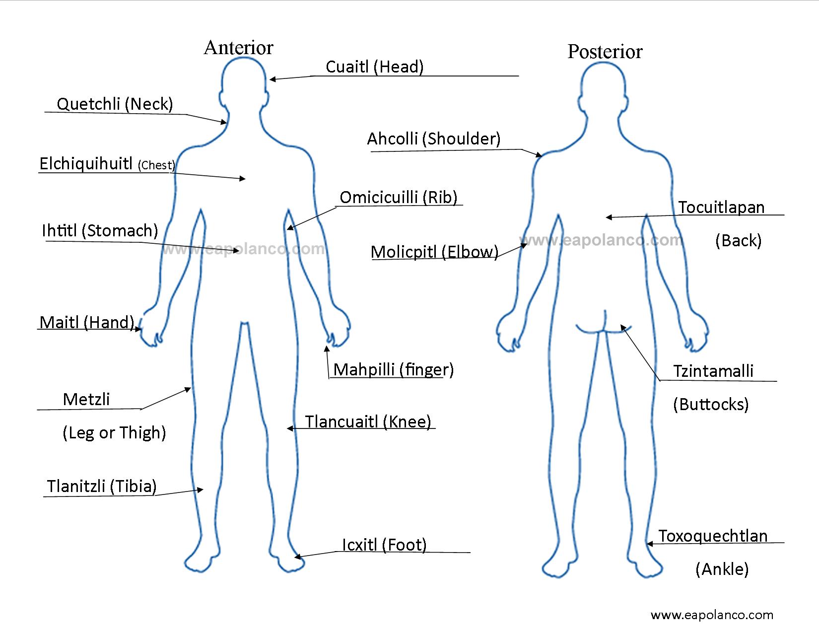
Body chart for medical diagram, front and back view. Blank unisex body outline template. Isolated vector illustration. concept of chiropractic technology or spine medical treatment, graphic of human back bone with x-ray interface Back muscles labeled anatomical educational body scheme vector illustration.

Muscle Facts Human Back Muscles DK Find Out
Human Body Diagrams INDEX Musculoskeletal Skeleton & Spine Shoulder & Back Arm & Hand Pelvis & Hip Leg & Foot Circulatory Nervous Digestive Urinary Reproductive Medical Art Library is a resource for teachers, students, health professionals or anyone interested in learning about the anatomy of the human body. We are medical artists who love anatomy.

Simple Muscle Chart Back Labeled Muscle Diagram Chart Free Download
Place your feet against the footrests with bent knees and grab your handle of choice. If you're unsure of which different row grip to choose, opt for a close-grip, neutral or overhand handle.

man lower back anatomy
The back is found posteriorly and includes the vertebral column, the muscles that support the back and the spinal cord. The vertebral column consists of 33 vertebrae which can be split up into 5 continuous sections. Each section is functionally different and is specialised for either weight-bearing, movement, protection and/or posture.

Back Muscles Diagram Female Back Muscles Anatomy Of Upper Middle
1 Diagrams 2 Human body diagrams 2.1 How to derive an image 2.1.1 Derive directly from raster image with organs 2.1.2 Derive "from scratch" 2.1.3 Derive by vector template 2.2 Examples of derived works 2.3 Licensing 3 Donating organs 4 Organs in other formats 4.1 Gastrointestinal 4.2 Other 4.3 Gastrointestinal Diagrams edit Human body diagrams

Lower Back Muscles Chart Diagram Of Back Muscles Of The Human Body
Anatomy Conditions Common injuries Treatments Summary The back supports the body's weight and allows for flexible movement while protecting vital organs and nerve structures. It comprises the.

Pin on Medical Assisting & Phlebotomy
Typical Anatomical Problems that Cause Back Pain. Spinal pain can arise from problems in the bones, nerves, or other soft tissues. Many of the intricate structures in the spine can lead to pain, and pain can be concentrated in the neck or back area, radiate to the extremities, or be referred to other parts of the body. For example:
human body back side parts
Your lower back contains 5 vertebral bones stacked above each other with intervertebral discs in between. These bones are connected at the back with specialized joints. The lumbar spine connects to the thoracic spine above and the hips below. Individual anatomical structures include 2 Cramer GD. The Lumbar Region.
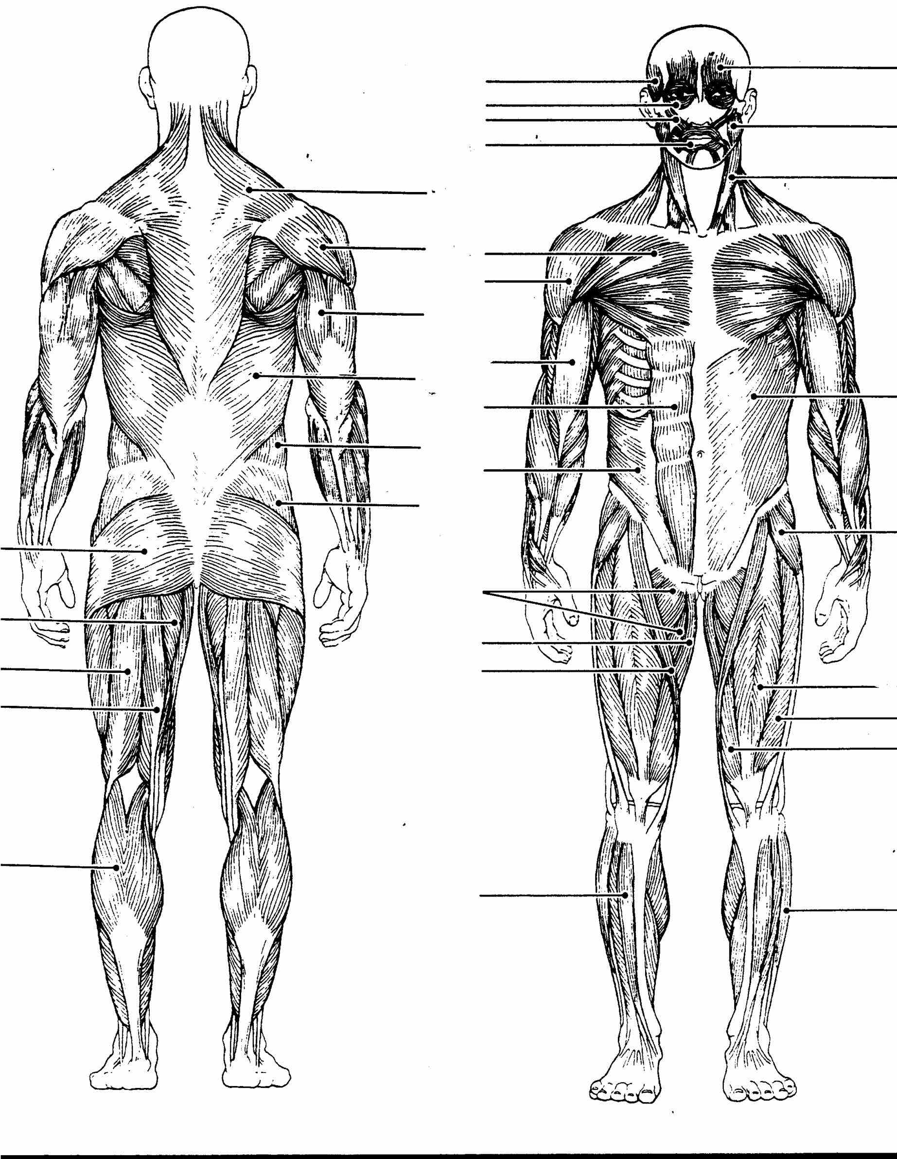
Major Muscles Of The Body Diagram
Anatomical diagrams of the spine and back These diagrams and original illustrations were produced from 3D medical imaging reconstructions of the spine and back by Micheua, Antoine - MD. All the images are in vector format, allowing an optimal web display with zoom and shifting of the anatomical images.
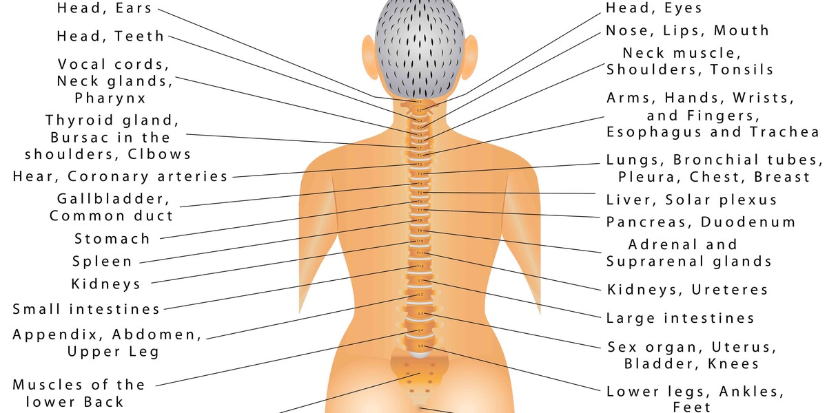
Diagram Of Common Back Bone Break Human Body Diagram / All of the
Back muscles diagram can also be considered to study the full location and shape of Latissimus Dorsi Muscle. Functions: Latissimus dorsi muscle is responsible for movements involving the arms.

Body Parts Diagram Of Human Body Organs Front And Back Pin On Human
What are your back muscles? Your back has many different muscles. Some muscles support your spine and trunk. Others help you move your body, stand up straight and assist with breathing. Because your back muscles support so much of your weight and are responsible for so many movements, injuries to these muscles are common.
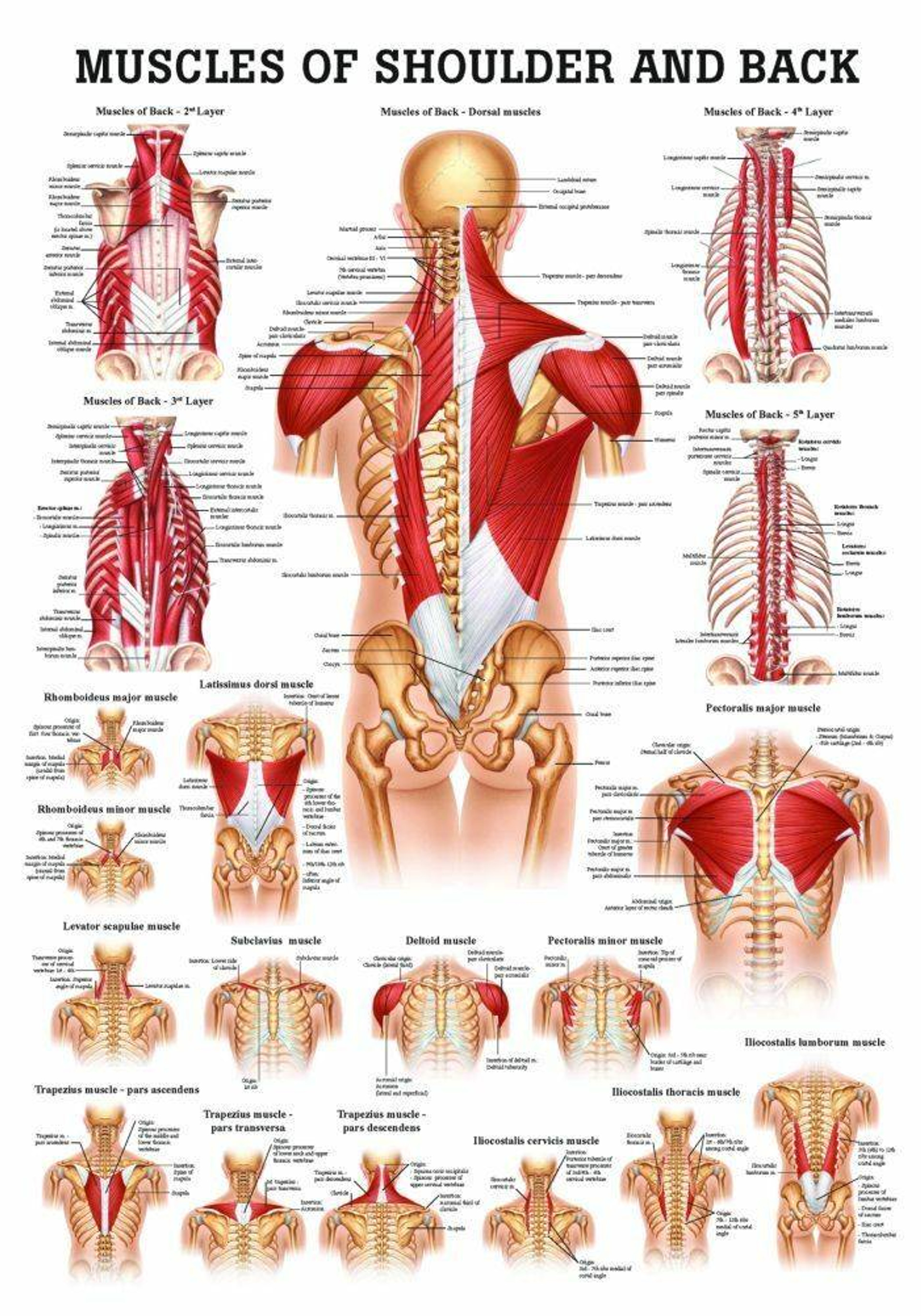
Muscles of the Shoulder and Back Laminated Anatomy Chart
Back Muscle Diagram With Lower Back Anatomy and Multifidus. This is a diagram of the larger and more surface muscles of the low back. The Multifidus muscles help to give segmental support to the spine.. The spasm of the muscles is your body's way of trying to protect the area that is hurting..

Printable Body Diagram Printable Word Searches
Your lumbar spine is the lower back region of your spinal column or backbone. It consists of five bones (L1-L5). Other structures in or around your lumbar spine are your intervertebral disks, spinal cord and nerves, muscles, tendons and ligaments. Your lumbar spine supports the weight of your body and allows a wide range of body movements.

Human Anatomy Diagram Organs Back View / The diagram shows five levels
Back anatomy The back is the body region between the neck and the gluteal regions. It comprises the vertebral column (spine) and two compartments of back muscles; extrinsic and intrinsic. The back functions are many, such as to house and protect the spinal cord, hold the body and head upright, and adjust the movements of the upper and lower limbs.

Back View Of Human Body Organs fx2design
Your back consists of a complex array of bones, discs, nerves, joints, and muscles. The muscles of your back support your spine, attach your pelvis and shoulders to your trunk, and provide mobility and stability to your trunk and spine. The anatomy of your back muscles can be complex. There are several different layers of muscles in your back.